[ad_1]
Successful drug development has a significant impact on the quality of life of people around the world. Being able to monitor how molecules enter target cells and observe what they do when they are inside is key to identifying the best candidates. Analytical techniques are therefore an important part of the drug discovery process. Researchers at Osaka University, in collaboration with RIKEN, reported a Raman microscopy-based approach for visualizing small molecule drugs using gold nanoparticles. The team’s findings are published in ACS Nano.
Small drug molecules are often tracked by connecting them to fluorescent probes that are visible when irradiated with light. Microscopy can then be used to see these molecules inside cells in real time. However, fluorescent molecules can be bulky, which can affect how small molecules behave. Additionally, some fluorescent molecules lose their fluorescence if they are exposed to too much light, making them difficult to see over the course of lengthy studies.
An alternative to fluorescent labels is a much smaller tag known as an alkyne, made up of triple carbon-carbon bonds. The particular arrangement of atoms in alkynes is not found naturally in cells; therefore, they provide a highly specific marker. Furthermore, their small size means that alkynes have a minimal effect on the behavior of small molecules. Instead of fluorescing under laser light, alkynes produce what is known as a Raman signal, which can be clearly identified among the cellular material signals.
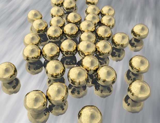
However, looking for the Raman signal of alkyne groups is complicated when there are not many around due to the low efficiency of Raman scattering. The researchers then combined alkyne labeling with the use of gold nanoparticles. Surface Enhanced Raman Diffusion Microscopy (SERS) can stimulate gold nanoparticles to produce enhanced electric fields that increase the Raman signal of alkynes, making them easier to detect.
“Our approach is a combination of techniques that have been used to trace small molecules in living cells,” says study lead author Kota Koike. “Gold nanoparticles are particularly useful messengers for signaling the presence of alkynes because they enhance the alkyne signal, as well as providing a surface that alkynes like to interact with. The two components then naturally join to generate the enhanced signal.”
Gold nanoparticles are readily absorbed by numerous different cell types, making the technique widely applicable. The nanoparticles enter the lysosome compartments within the cell and thus enhance the signal of the alkyne-labeled molecules that subsequently arrive in the lysosomes and interact with them.
Our SERS technique has the potential to be used with a variety of different cell types and with a virtually unlimited number of drug candidates. This is particularly interesting for drug discovery, where any means to better understand drug dynamics in real time is extremely valuable for development. “
Katsumasa Fujita, corresponding author of the study
Source:
Journal reference:
Koike, K., et al. (2020) Quantitative dynamics of drugs visualized by plasmon-optimized Raman microscopy marked by alkynes. ACS Nano. doi.org/10.1021/acsnano.0c05010.
.
[ad_2]
Source link
