[ad_1]

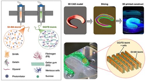
A highly elastic hybrid construct for fibrocartilage regeneration is produced by covering a gellan gum composite bioink loaded with cells / fibrinogen together with a silk methacrylate fibroin bioink in a cross hatch pattern. Credit: WFIRM
Scientists at the Wake Forest Institute for Regenerative Medicine (WFIRM) have developed a method to bioprint a type of cartilage that could one day help restore knee function damaged by arthritis or injury.
This cartilage, known as fibrocartilage, helps connect the tendons, ligaments or bones and is found mainly in the meniscus of the knee. The meniscus is the hard, rubbery cartilage that acts as a shock absorber in the knee joint. Meniscal tissue degeneration affects millions of patients and arthroscopic partial meniscectomy is one of the most common orthopedic operations performed. In addition to surgery, there is a lack of available treatment options.
In this latest proof-of-concept strategy, scientists were able to 3-D bioprint a hybrid tissue construct for cartilage regeneration by printing two specialized bioinks – hydrogels that contain cells – together to create a new formulation that provides a friendly microenvironment cell and structural integrity. This work is done with the Integrated Tissue and Organ Printing System, a 3-D bioprinter developed by WFIRM researchers over a period of 14 years. The system deposits both biodegradable materials, plastics to form the “shape” of tissue and bioinks that contain cells to build new tissues and organs.
“In this study, we were able to produce a highly elastic hybrid construct for advanced fibrocartilage regeneration,” said Sang Jin Lee, Ph.D, associate professor at WFIRM and author of the paper recently published by Chemistry of materials magazine. “The results demonstrate that this bioprinted construct offers a versatile and promising alternative for the production of this type of tissue.”
For the study, Lee and the WFIRM research team tested various formulations and measured the response to applied forces or stresses, the swelling ratio, and the strength and flexibility of the material. One provided the appropriate cellular microenvironment to maintain cells and help them grow, while the other bioink offered excellent biomechanical behavior and structural integrity. The final formula of the two bioinks used was co-printed layer by layer to create a mesh pattern. The constructs were implanted in a small animal model for observation for 10 weeks and evaluated at intermittent time periods, showing proper functioning.
“A larger preclinical study will be needed to further examine the body’s response and functional joint recovery with the use of this regenerative medicine treatment,” said James Yoo, MD, Ph.D., professor at WFIRM.
“We have such a need for effective treatments and therapies to help patients cope with degenerative joint problems, particularly the knee,” said Anthony Atala, MD, director of WFIRM. “This proof-of-concept study helps steer our work in the right direction to one day be able to design this crucial tissue that is so important to patients.”
Neural cells accelerate function in 3-D bioprinted skeletal muscle constructs
João B. Costa et al, 3D bioprinted highly elastic hybrid constructs for advanced regeneration of fibrocartilage tissue, Chemistry of materials (2020). DOI: 10.1021 / acs.chemmater.0c03556
Provided by Wake Forest University Baptist Medical Center
Quote: Scientists Create Hybrid Tissue Construct for Cartilage Regeneration (2020, Nov 9) Retrieved Nov 9, 2020 from https://phys.org/news/2020-11-scientists-hybrid-tissue-cartilage-regeneration. html
This document is subject to copyright. Aside from any conduct that is correct for private study or research purposes, no part may be reproduced without written permission. The content is provided for informational purposes only.
[ad_2]
Source link
