[ad_1]
DURHAM, NC – The bacterium that causes tularemia, the tick-borne disease, is a lean and mean infecting machine. It carries a relatively small genome and a unique set of infectious tools, including a collection of chromosomal genes called the “island of pathogenicity”.
A team of researchers from Duke University, Harvard, the University of Rhode Island and the National Institutes of Health have now unpacked the bacterium’s toolbox and built an understanding of the shapes and interactions of all its parts, using a technique imaging technique called cryo-electron microscopy.
Their insights, which appear Nov. 19 on Molecular Cell, point to one way the bacterium’s unique infectious mechanism could be blocked.
The Francisella tularensis bacterium can infect more than 200 types of animals including humans, dogs, cats, fish and rodents. A minimum of ten bacteria, usually from an animal or insect bite, are enough to be an infectious dose. Tularemia attacks the skin, eyes, lymph nodes and lungs, sometimes leading to pneumonia, meningitis, inflammation around the heart, and bone infections. Fortunately, it’s rare in humans, but it can be deadly and make people sick.
In fact, since the 1920s, Russia, the United States and other countries have studied it as a potential biological weapon.
The bacterium is capable of infecting many different types of cells, but its weak point is the macrophage, an immune system cell that is a first responder to invading bacteria.
When an alien bacterium is detected, a macrophage will envelop the insect and capture it inside a bubble in its cell membrane called a phagosome, where it will be chewed and rendered impotent. It is usually the end of the story.
But Francisella, sensing that he has been captured, turns on a specialized set of his own genes, the island of pathogenicity, and begins to create the tools he will need to escape the phagosome and enter the fluid center of the macrophage, where it can replicate in peace. .
“It’s a kind of professional pathogenic bacterium,” said Maria Schumacher, a distinguished professor of biochemistry at the Duke School of Medicine, who is a co-correspondent author of the study. One of the key parts of its infectious machinery is a protein called MglA (macrophage growth locus protein A), which has not been found anywhere else. “This bug developed these special proteins,” Schumacher said. “It is also very robust; it can survive even in dead carcasses and soil. “
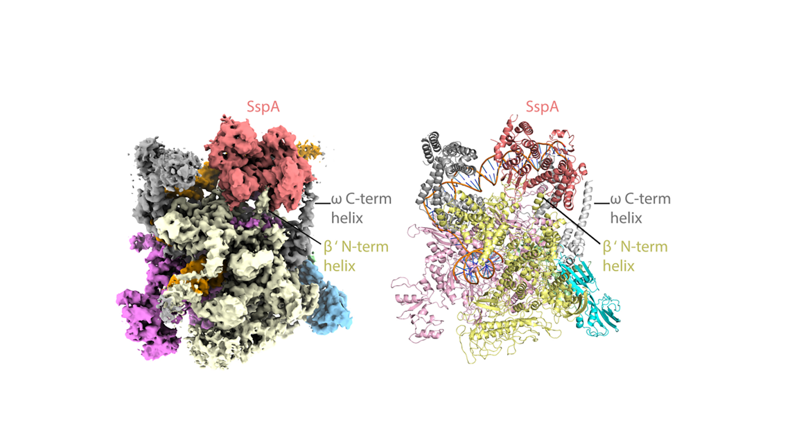
The researchers used state-of-the-art cryo-EM microscopes at Duke and Harvard to see the shapes and interactions of all of the bacterium’s infectious machinery and how they worked together.
The researchers were able to determine the shapes and interactions of all the players in the system, from the small molecule signal to the large multiprotein complex, a “specialized virulence RNA polymerase” that activates the genes of the pathogenicity island.
“It’s a new system and that’s why we’re so excited about it,” said Schumacher. “It is not like any other that we have found in any other bacterium or eukaryote. It is all very unique and interesting. “
“Even if you know all of these components, how they work together is not clear,” Schumacher said. “Could we actually look at it and see how it works?” Key structural analyzes on large molecular complexes were performed by Brady Travis, a graduate student in biochemistry in the Schumacher laboratory and first author of the study, who mastered cryo-EM to enable visualization of these complexes.
Schumacher and corresponding co-author Richard Brennan, president of biochemistry at Duke, are now pursuing a grant to screen a collection of small molecules that could bind to MglA and prevent it from allowing the assembly of this specialized virulence complex.
“We found a nice pocket for binding potential inhibitors of MglA function,” Brennan said. “If you can keep him from bonding with his partner, you won’t get the pathogenic island transcript, which is a good thing.”
This research was supported by the National Institutes of Health, the Department of Energy and the US National Science Foundation. (R35GM130290, R21AI146641, AI081693, AI145954, F31AI150138, HD055148-08, U24GM129547, DE-AC02-05CH11231, ECCS-1542015)
QUOTE: “Structural Basis for Virulence Activation of Francisella tularensis,” Brady A. Travis, Kathryn M. Ramsey, Samantha M. Prezioso, Thomas Tallo, Jamie M. Wandzilak, Allen Hsu, Mario Borgnia, Alberto Bartesaghi, Simon L. Dove, Richard G. Brennan, Maria A. Schumacher. Molecular Cell, November 19, 2020 DOI: 10.1016 / j.molcel.2020.10.035
.
[ad_2]
Source link
