[ad_1]

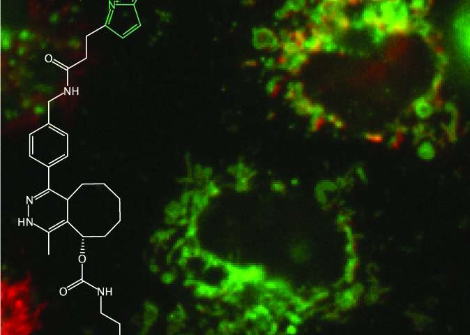
Universal single residue terminal labels for imaging fluorescent live cells of microproteins. Credit: Simon Elsasser
The SciLifeLab Simon Elsässer laboratory at the Karolinska Institutet reports a method that allows for the fluorescent labeling of proteins with the small perturbation – a single amino acid – genetically added to both ends of a (micro) protein of interest. The method is called Single-residue Terminal Labeling (STELLA).
Thirty years ago, the cloning of the green fluorescent protein GFP, along with genetic engineering tools, revolutionized the field by allowing researchers to fuse a fluorescent “beacon” to any protein of interest so that it can be observed directly in living cells using fluorescence microscopy. Today’s microscopes obtain live, nanometer-resolution, multicolor images, allowing researchers to resolve even the smallest subcellular structures. However, fluorescent proteins have a limitation: the size of the fluorescent tag is often equivalent to the size of a typical folded protein, thus adding a considerable molecular “load” to the protein under study and potentially affecting its function. This can become a particular obstacle to the study of microproteins, a recently appreciated class of proteins and much smaller than average.
In a study by postdoctoral researcher Lorenzo Lafranchi of the Simon Elsässer laboratory at the Karolinska Institutet SciLifeLab, a reported method, which allows fluorescent labeling of proteins with the small perturbation – a single amino acid – added genetically on both ends of a (micro) protein of interest. The method is called single residue terminal labeling, STAR. It is based on a synthetic building block (a non-canonical “designer” amino acid, rather than one of the canonical 21) that is incorporated together with a larger tag using a technique called expansion of the genetic code. The tag is then rapidly removed from the cell, leaving a single terminal designer amino acid on the protein of interest. The designer amino acid introduces a chemical group into the protein that subsequently allows conjugation with a small organic fluorescent dye, illuminating the protein of interest within the living cell. The advantage over existing labeling techniques that rely on the expansion of the genetic code and STELLA can be used to label the terminals of any protein.
The study, published in Journal of the American Chemical Society, demonstrates the usefulness of STELLA in the fluorescent labeling of a variety of human proteins and microproteins, located in different subcellular compartments and organelles. In addition to cellular proteins, the team was also able to label and locate a series of elusive polypeptides produced by the SARS-CoV2 coronavirus that causes Covid-19.
Illumination technique for cell surface receptors developed by researchers
Lorenzo Lafranchi et al, Universal Single-Residue Terminal Labels for Fluorescent Live Cell Imaging of Microproteins, Journal of the American Chemical Society (2020). DOI: 10.1021 / jacs.0c09574
Provided by Karolinska Institutet
Quote: Illumination of Tiny Proteins in Living Cells Using Single Residue Labeling Tags (2020, November 12) Retrieved November 12, 2020 from https://phys.org/news/2020-11-illuminating-tiny-proteins-cells -single-residue. html
This document is subject to copyright. Aside from any conduct that is correct for private study or research purposes, no part may be reproduced without written permission. The content is provided for informational purposes only.
[ad_2]
Source link