[ad_1]
Scientists have developed an optical elastography technique that could revolutionize the accuracy and ease with which healthcare professionals can detect biomechanical changes in cells and tissues.
A study derived from an international collaboration between the University of Exeter, the Gloucestershire Hospitals NHS Foundation Trust, the University of Perugia (Italy) and the Institute of Materials of the National Research Council of Italy (IOM-CNR) applied an approach innovative biophotonic to highlight how microscopic processes drive mechanical modification in biological tissues.
The team of experts, coordinated by Dr. Francesca Palombo of the University of Exeter and Prof. Daniele Fioretto of the University of Perugia, Italy, analyzed the great potential of the technique in microscale tissue investigation.
While the mechanical properties of both cells and tissues play a vital role in cell function and how disease develops, traditional methods of studying these properties can be limited and invasive.
Scientists recently used Brillouin microscopy, a form of imaging that uses light to create an acoustic measurement of cells and tissues, as a method for non-invasive studies of these biomechanical properties.
However, a complicating factor in these measurements is the contribution of water to both the biomechanics of tissues and cells, and to the Brillouin spectrum itself.
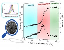
Now, for the new study, the team used natural biopolymer hydrogels to mimic human tissue and compare the results with measurements made in human tissue samples.
They found that this new technique allows for the study of functional properties (and alterations) of tissues at the subcellular scale, which means that practitioners can gain insight from the analysis of a new space-time region of biological processes.
The results of this study show that, while water plays an important role in determining mechanical properties, the effect of the solute, including proteins, lipids and other components, is most evident on viscosity, which is relevant for transport. metabolites and active molecules.
The research was published in Science Advances.
Dr Palombo, associate professor in biomedical spectroscopy at the University of Exeter, said, “We set out to understand the basics of Brillouin signals in biomedical samples.
“While taking a step back to analyze the fundamentals of this light scattering process, we have made substantial progress as we now understand the distinctive contribution of interfacial dynamics, beyond bulk water, to the viscoelastic response of biological tissues.
“This has far-reaching implications as phase changes, as well as acoustic anisotropy, are ideal scenarios in which Brillouin imaging provides unique information. We are still working to establish the relevance of this technique in the medical sciences, however it is indisputable that it offers an invaluable contrast mechanism to detect physiological and pathological states “.
Viscoelastic properties of biopolymer hydrogels determined by Brillouin spectroscopy: a tissue micromechanics probe is published in Science Advances.
.
[ad_2]
Source link