[ad_1]
Coronaviruses are a group of RNA viruses that cause disease in mammals and birds. In humans and birds, they cause respiratory tract infections that can range from mild to fatal. Mild illnesses in humans include some cases of the common cold. Conversely, more lethal varieties can cause SARS, MERS and now COVID-19 disease. The newest type of coronavirus, severe acute respiratory syndrome coronavirus-2 (SARS-CoV-2), along with the earlier SARS-CoV-1 and MERS-CoV, fall into the category of beta coronaviruses. There have not yet been any noteworthy developments in the therapeutic approach for these beta-type CoVs, which makes it interesting to design and develop a unique therapeutic pathway to minimize transmission of SARS-CoV-2 and the impact of COVID- 19.
COVID-19 disease caused by the SARS-CoV-2 virus causes a wide range of diseases starting from mild respiratory disease, severe progressive pneumonia and acute respiratory distress syndrome, increased cytokines, multi-organ failure and death. Neutrophils, which make up about 70% of the white blood cells in our body, are highest in the severe progression of COVID-19 disease. Abnormal activation of neutrophils triggers the release of extracellular neutrophil traps (NETs) and extracellular DNA leading to multi-organ failure and septic shock.
Because the treatment of severe COVID-19 is very difficult, researchers from the Korea Research Institute of Biosciences and Biotechnology (KRIBB) have suggested that DNase-1 can be used to dissolve neutrophil extracellular traps (NETs), thus stopping the further progression of COVID-19. Since DNase has a short half-life in blood plasma, Wonha Lee and colleagues proposed coating the DNase on the surface of the nanoparticles to stabilize and preserve DNase activity. Researchers used polydopamine to attach DNase to the surface of the nanoparticles. The research is published in the journal Advanced Sciences.
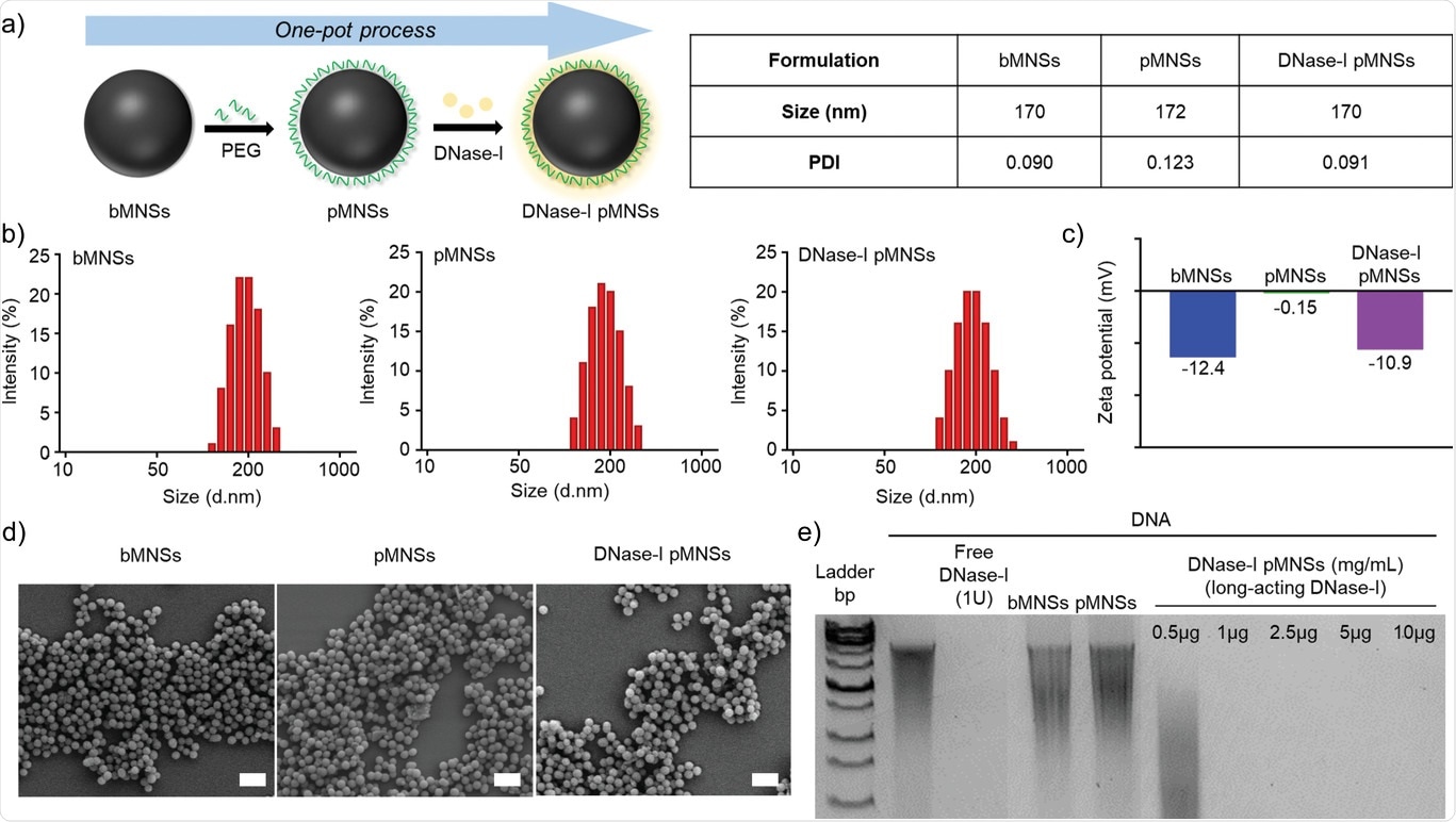
 Preparation of DNase-I pMNS. b) Size distribution of bMNS, pMNS and DNase-I pMNS. c) Zeta potentials of bMNS, pMNS and DNase-I pMNS. d) Scanning electron microscope (SEM) images of bMNS, pMNS and DNase-I pMNS (scale bar: 500 nm). e) Migration profile of pure DNA after digestion with free DNase - I, pMNS and bMNS, as well as various amounts of pMNS DNase - I. pMNS, melanin-like nanospheres coated with PEG; bMNS, nude bio-inspired nanospheres similar to melanin.")
Physico-chemical characterization of pMNS. a) Preparation of DNase-I pMNS. b) Size distribution of bMNS, pMNS and DNase-I pMNS. c) Zeta potentials of bMNS, pMNS and DNase-I pMNS. d) Scanning electron microscope (SEM) images of bMNS, pMNS and DNase – I pMNS (scale bar: 500 nm). e) Migration profile of pure DNA after digestion with free DNase – I, pMNS and bMNS, as well as various amounts of pMNS DNase – I. pMNS, melanin-like nanospheres coated with PEG; bMNS, nude bio-inspired nanospheres similar to melanin.
Development of the new treatment strategy for COVID-19
Initially, the researchers analyzed computed tomography of severely infected COVID-19 patients whose lungs were severely damaged. They found that the patients had higher neutrophil counts and lower lymphocyte counts, another type of white blood cell responsible for fighting infection. They also compared extracellular DNA, DNAse, and histone citrullinate H3 in healthy and COVID-19 infected patients. Lee found that extracellular DNA and histone H3 citrullinate levels were increasing in severely infected individuals, while DNase-I levels were decreasing in infected patients.
Next, the scientists synthesized melanin as nanoparticles using dopamine oxidation. The surface of the melanin-like nanoparticles was immobilized with DNAse – I and polyethylene glycol (PEG). Visualization of synthesized and coated nanoparticles under a scanning electron microscope revealed that the morphology of the nanoparticles was spherical and had a uniform size distribution. Researchers evaluated the degradation ability of DNAse-I and PEGylated nanoparticles and found that the DNA was degraded to a nanosphere concentration greater than 1 microgram. They also evaluated the efficiency and stability over time of DNAse-I binding on nanoparticles through the BCA assay and treatment with FBS with PBS and PBS media only. As DNAse-I concentration increased, binding efficiency also increased and the DNASe-I-coated nanospheres remained stable for up to 72 hours with PBS media alone.
Effect of DNAse-I PEGylated melanin-like nanoparticles on COVID-19 patients
Severely affected COVID-19 patients suffer from a decline in DNAse-I in their system, requiring the use of PEGylated DNAse-I melanin-like nanoparticles. The team of scientists from the Korea Institute of Biosciences and Biotechnology treated plasma from COVID-19 patients and surprisingly found that both free DNAse and DNAse coated nanoparticles reduced levels of extracellular DNA. Furthermore, they found that factors such as myeloperoxidase, the neutrophilic elastase responsible for the extracellular traps of neutrophils that consequently increase the severity of COVID-19 were also alleviated to a greater extent when treated with DNAse-I PEGylated melanin-like nanospheres. More than half of the COVID-19 patients admitted to hospital had sepsis caused by uncontrollable inflammatory responses. Inflammation is characterized by an increase in cytokine levels. They quantified the immune factors responsible for inflammation after treatment with DNase-I and DNAse-I-coated nanoparticles and found that the factors were significantly reduced, promising that these DNAse-coated nanoparticles can be used as a potential therapeutic approach.
Effect of nanoparticles on septic mouse model treated with caecal ligation and perforation (CLP)
Researchers further validated PEGylated DNase-I-synthesized nanoparticles in vivo using a CLP mouse model. To create a CLP model, the opening of the large intestine called the cecum is opened and holes are made to induce inflammation, mimicking sepsis in humans. The KRIBBS scientists administered DNase-I-like melanin nanospheres on the CLP mouse model and found that the number of neutrophils dropped dramatically. Scientists also realized a 40% survival rate and reduction in cytokine levels in CLP models after administration of nanoparticles, confirming that these nanoparticles can be used as a potential treatment strategy.
Conclusion
Researchers have shown that extracellular DNA, one of the factors responsible for the progression of COVID-19, can act as a likely target for the treatment of SARS-CoV-2 induced sepsis. They also found that the release of PEGylated melanin-like nanospheres with DNase I suppressed extracellular DNA expression, which in turn slowed the progress of sepsis in severe COVID-19 patients. They say early injection of these nanoparticles can potentially reduce the progression of COVID-19 disease. Although the efficiency of bioinspired nanospheres in both in vitro blood sample testing and in vivo murine septic model testing is verified, Lee recommends validation of the nanoparticles under appropriate in vivo sepsis-induced animal models prior to proceeding to clinical trials on Human COVID-19 patients. That said, PEGylated melanin-like nanospheres could be a promising therapeutic approach for SARS-CoV-2 sepsis.
.
[ad_2]
Source link
