[ad_1]

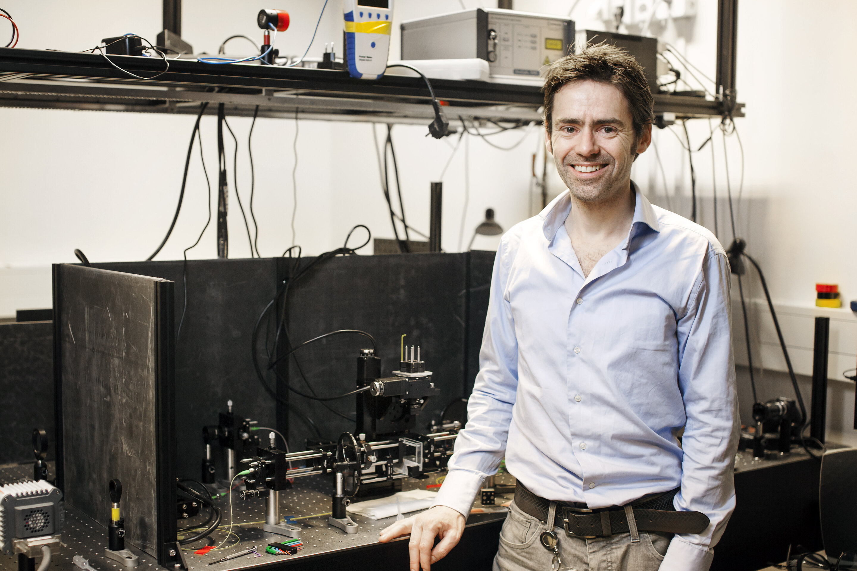
Researcher Jeroen Kalkman is alongside his new imaging setup. Credit: TU Delft
One of the challenges in optical imaging is to visualize the interior of the tissue in high resolution. Traditional methods allow researchers to look at a depth of about 1 millimeter. Researchers from the Delft University of Technology have now developed a new method that can penetrate up to four times deeper, up to about 4 millimeters. The healthcare sector, in particular, could benefit from the new technique in the future.
The new imaging method brings together a number of existing techniques. The most important of these is optical coherence tomography, a technique used by ophthalmologists to visualize the retina. OCT is similar to acoustic ultrasound but uses light instead of sound waves while having a higher resolution. Using the information contained in the reflected light waves, an algorithm can create a cross section of the tissue.
Cross section
Unlike a normal OCT scan, the Delft researchers do not make images with reflected light, but send the light directly through the tissue. On the other hand, a sensor captures it again. Researchers can see what light comes in and when. “Light that travels over a longer period of time is scattered through the tissue and arrives at the detector relatively late,” explains TU Delft researcher Jeroen Kalkman. “Usually, this causes the resulting images to be blurry. But by looking at the time of arrival, we can separate this scattered light from the light that passed directly through the sample. With the light arriving early, we can produce an image clear. “
To make a cross section, a so-called tomogram, of the object, the researchers use technologies known from computed tomography, of which the best known example is the CT scan. “This involves measuring a projection of the X-rays that pass through the object from many different angles and positions,” says Kalkman. “You can then link all these different projections together using a computer to create a three-dimensional image. We do the same thing, but with light.”
To find out how powerful their technique is, the researchers tested it on dead zebrafish, which they obtained through an ongoing study at Erasmus MC. The maximum depth of penetration was found to be approximately four millimeters, a factor of four improvement over the current reflection approach in OCT. Furthermore, the organs of the zebrafish could be represented in high contrast by observing both the strength and the time of arrival of light. Kalkman says, “We’ve been working on this with a whole team of researchers for nearly ten years, so it’s a huge thrill that we finally did it.”
In the future, the new Delft technique could generate valuable information on some diseases. “With our method, we would be able to track the development of such a disease very precisely over time,” says Kalkman. “In this way, we could study the effects of drugs or, conversely, potentially toxic substances on tissues. This could provide us with useful information that can ultimately lead to better treatments or better protection.”
Another application of the new method is the analysis of biopsies, small pieces of human tissue that doctors take from patients for analysis. “Currently, laboratories often add fluorescent labels to biopsies, or cut them into small slices and use optical compensation to make them more transparent,” says Kalkman. “This takes a long time and during this process the biopsies can deform. We expect our technique to be able to visualize the biopsies in their three-dimensional form, thus helping doctors to make a more accurate diagnosis.”
3-D optical biopsies at your fingertips thanks to advances in light field technology
Unlabeled Quantitative Optical Deep Tissue Tomography, Jelle van der Horst, Anna K. Trull, Jeroen Kalkman, Optics. www.osapublishing.org/optica/f… -7-12-1682 & id = 444004
Provided by the Delft University of Technology
Quote: Researchers peer deep inside tissues with new high-resolution techniques (2020, December 1) recovered December 1, 2020 from https://phys.org/news/2020-12-peer-deep-tissue- high-resolution-techniques.html
This document is subject to copyright. Aside from any conduct that is correct for private study or research purposes, no part may be reproduced without written permission. The content is provided for informational purposes only.
[ad_2]
Source link
