[ad_1]
Most vaccine research in the current 2019 coronavirus (COVID-19) pandemic has focused on the role of the angiotensin converting enzyme 2 (ACE2) receptor in mediating viral entry into the host cell. However, a recent study published on the prepress server bioRxiv* in November 2020 discovers the major role played by the N-terminal domain of the SARS-CoV-2 virus in host infection. This could be of great use in measuring the usefulness of antibodies used in convalescent plasma therapy for COVID-19.
Until now, the virus spike protein (or protein S) has been observed to interact with the host cell ACE2 receptor, with the S1 subunit involved in virus receptor binding via the receptor binding domain (RBD). RBD is now universally recognized as the primary target site for neutralizing antibodies.
However, RBD is structurally linked to the N-terminal (NTD) domain of the adjacent spike protomer and monoclonal antibodies against NTD neutralize the infection, suggesting that NTD, as well as RBD, play a pivotal role in SARS-CoV -2 infection. “

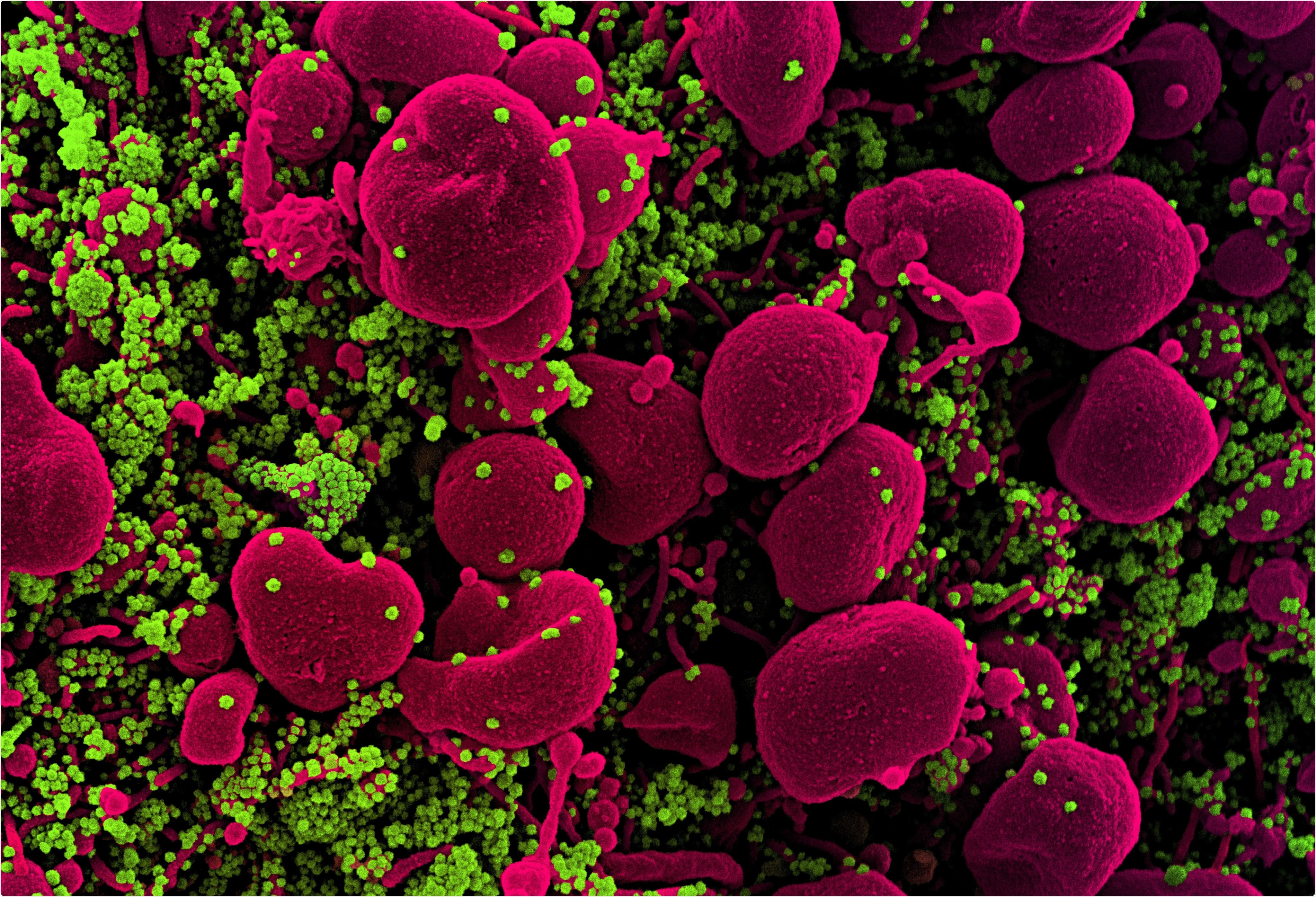
Study: The N-terminal domain of spike glycoprotein mediates SARS-CoV-2 infection by associating with L-SIGN and DC-SIGN. Image credit: NIAID
Evidence of support for the role of the NTD
Although COVID-19 is primarily a respiratory infection, with the main complication in severe cases of pneumonia, ACE2 is not highly expressed in lung cells, occurring at relatively low levels in type II pneumonocytes. This has caused understandable confusion, in addition to which recent findings have shown that cells that do not express ACE2 also contain SARS-CoV-2 RNA. Therefore, there may be other receptors that allow the virus to enter specific organs such as the lungs.
Researchers from Osaka University, Japan used a pulmonary cDNA library to identify potential receptors, as well as the mechanism by which the virus successfully spreads to other organs such as intestines, liver, kidneys and blood vessels in infected patients. .

Lung cDNA library screening using SCoV2 spike NTD-Fc. a, The pulmonary cDNA library screening workflow. b, cells transfected with the retrovirus cDNA library were stained with SCoV2-NTD-Fc. Proportions of cells stained with SCoV2-NTD-Fc are shown. c, Agarose DNA gel electrophoresis of genes derived from amplified cDNA libraries from a single cell clone stained with SCoV2-NTD-Fc. The indicated band has been identified as L-SIGN. d, The clone of a single cell stained with SCoV2-NTD-Fc was analyzed by mAb anti-DC-SIGN and mAb anti-DC / L-SIGN (red line) or control (gray gradient).
L-SIGN acts as an NTD receptor
The researchers found that in all cases, cells containing the viral spike protein NTD also contained the L-SIGN receptor and were stained with anti-L SIGN antibodies. This indicates, they say, “that L-SIGN is one of the major 86 molecules that interacts with SCoV2-NTD in the lung.” In particular, this receptor is highly expressed in the lung, despite its main site of expression being the liver.
On the other hand, none of the cells contained the RBD.
NTD binds to L-SIGN / DC-SIGN via unique glycans
L-SIGN and its closely related DC-SIGN receptor are both found in the lung. Another receptor called CD147 is suspected of mediating SARS-CoV-2 infection. However, the researcher found that only NTD bound to both L-SIGN and DC-SIGN on the cell surface, but not to ACE2 or CD147.
It was found that RBD only binds to ACE2. Again, both L-SIGN and DC-SIGN are ligands of the intercellular adhesion molecule 3 (ICAM-3), via a sugar chain. Sugar called mannan is a ligand for both of these receptors, and its presence greatly suppresses the binding of NTD to one of the aforementioned receptors. The researchers comment: “The unique glycans on NTD of SARS-CoV-2 on N149 are involved in the interaction with L-SIGN and DC-SIGN. “
The unique interaction between the SARS-CoV-2 NTD peak and these receptors is mirrored by the failure of the SARS-CoV NTD, or the other human coronaviruses OC43 and HKU1, to do the same.
SIGN receptors mediate membrane fusion
Membrane fusion is essential for successful enveloped virus infection. The researchers found that cell-to-cell fusion occurred when cells infected with the viral spike protein were grown alongside cells that expressed the SIGNS on their surface. This indicates that these receptors cause membrane fusion once the cell that expresses them is the target of the virus.
Again, the researchers found that DC-SIGN, which was expressed on monocyte-derived DCs (moDCs), but SARS-CoV-2 cannot produce membrane fusion in the presence of the CD74 receptor. Therefore, SIGNs expressed in CD74-negative cells are likely responsible for mediating this infection.
Second, since viral NTD binds to moDCs, it is possible that the virus attached to DC-SIGN on moDCs could infect other cells, as shown in a co-culture experiment. And indeed, not only is L-SIGN expressed in lung and endothelial cells, but on DCs and specialized macrophages that carry DC-SIGN, and which are also found in the lung.
The researchers comment: “Their localization in the lung further strengthens the increasingly likely role of L-SIGN and DC-SIGN as SCoV2 receptors and may therefore be important in the pathogenesis of pneumonia.”
This infection was stopped in vitro from anti-DC-SIGN antibodies or from mannan glycan. The prompt spread of the virus can be explained by this phenomenon since DCs are circulating cells.
Anti-NTD / RBD antibodies neutralize SIGN-mediated SARS-CoV-2 infection
Both anti-RBD and anti-NTD antibodies blocked the infection by the SARS-CoV-2 pseudovirus on SIGN-carrying cells, showing the ability of serum antibodies to neutralize viral infection through these receptors and indicating that both the receptors play a role in successful infection.
Implications and future directions
The study therefore sheds some light on the possibility that SARS-CoV-2 may act through other host receptors, particularly SIGNs, to achieve infection of non-ACE2-carrying cells and dissemination to non-pulmonary organs.
Second, the interaction between the NTD of this virus and the SIGNs seems unique to the current virus since neither the previous SARS virus (severe acute respiratory syndrome) nor seasonal coronaviruses show efficient binding of spikes to these receptors. In fact, the SARS-CoV-2 spike protein has several glycan groups attached to it than the others, even SARS closely related. Furthermore, the glycan N bound to residue 149, which is mainly responsible for the NTD-DC-SIGN interaction, is absent in the latter and contains a pentasaccharide motif (GlcNAc2Man3) recognized by the SIGNs.
Thirdly, the spread of the virus throughout the body and the occurrence of serious complications, including thrombosis, can be explained by the presence of these receptors on endothelial cells and many other organs in the body. Investigators explain: “Because of their migration through the body, DCs can function as transporters [of SARS-CoV-2] associating with the virus via DC-SIGN to infect cells that express ACE2 or L-SIGN, which in turn facilitates the spread of the virus into the host. “
Again, the presence of neutralizing antibodies found in COVID-19 patients targeting SARS-CoV-2 infection by DC-SIGN receptors indicates that NTD is a therapeutic target. It is important to note that most of the monoclonal antibodies from these patients fail to bind to RBD and that neutralizing antibodies do not prevent spike-ACE2 binding. However, the anti-RBD 4A8 antibody prevented infection by the virus of the SIGN carrier cells. Therefore, the study supports the importance of using convalescent plasma for the treatment of these patients, in particular antibodies directed against NTD.
The study concludes: “Further analysis of L-SIGN or DC-SIGN-mediated infection would be important for understanding the etiology of SARS-CoV-2 related diseases. Furthermore, targeting SIGN-mediated infection and ACE2-mediated infection would be important for effective vaccine development. “
*Important Notice
bioRxiv publishes preliminary scientific reports that are not peer-reviewed and, therefore, should not be considered conclusive, guide clinical practice / health-related behavior, or treated as consolidated information.
source
.
[ad_2]
Source link